


Cangzhou Pangoo International Trade Co., Ltd.
News
- 5 ways to help you choose high quality probiotic products1. Strains: strains are very important for the production of probiotic products. Strains detecting is a long process.Technical requirements are relatively high. The powerful manufacturers have their own laboratories and research and development staff, who can detect the strains are good or bad. 2.Activity: the product is very effective, while you start to use it at the beginning. But after a month the effect is not good. Just because the viability of the product is not so high. Live bacteria has been damaged during the production process in small factories, therefore losing the ability to reproduce . From our customers` feedbacks, we get to know the activity of pangoo products is very good, and resistant to storage. We have tested the Biobed which are kept for a year and found that the loss rate is only 10%. This loss rate is within error allowance, so there is almost no loss. 3.Adding proportion: live bacteria with high purity produced by the powerful factory. The adding ratio is 0.1%-0.2%. At the same time, the single strain is difficult to play its role . The compound product is made of a variety of beneficial living bacteria. This kind of product is made by special mixing. Therefore, the strains between the compound can do mutual promotion,gaining a better effect. 4.Technology: Three technologies are applied at present: they are solid fermentation, liquid fermentation, liquid – solid fermentation. Through Liquid fermentation, a lot of live bacteria and spore can be produced,less infectious microbe. However,this technology cost a lot. It is usually applied in medicine. Solid fermentation produces low number of live bacteria and spores with more infectious microbe . but the activity of bacteria is more stable and easier to store. Liquid – Solid fermentation technology is adapt by pangoo.Through this technology , a large number of live bacteria with high stability can be produced, At the same time, cost can be controlled. 5. Package: most microbial agents are sensitive to the oxygen in the air. The packing should choose well sealed and moisture-resistance materials, such as good aluminum foil or high quality seal non-toxic plastic calabash.
2017 10/18
- Composting Chicken Manure1.Introduction Composting chicken manure is a side benefit of raising chickens. Chicken manure fertilizer is very high in nitrogen and also contains a good amount of potassium and phosphorus . But the high nitrogen in the chicken manure is dangerous to plants if the manure has not been properly composted. There are various ways of processing chicken manure, such as physical methods, chemical methods, microbial fermentation methods. The traditional way of making fertilizer by chicken manure is to naturally accumulate it to for 5 to 6 months`fermentation . In such way ,the smell not only causes air pollution but also causes nutrient loss. However , If the chicken manure are treated by microbial fermentation , it can not only speed up the fermentation process, shorten the fermentation time, prevent waste to improve the nutritional value ; but also dry chicken manure by temperature produced byfermentation, eliminate odor, reduce environmental pollution. In the fermentation of chicken manure,The main microorganisms that play a key role are Bacillus subtilis, Bacillus licheniformis, Candida albicans, Candida utilis, Bacillus megaterium, Bacillus licheniformis, Lactobacillus plantarum, Feces Enterococcus, Trichoderma reesei, protease, cellulase and other beneficial microorganisms. These flora in the growth of metabolic substances produced in each other. In the process of fermentation, microbial flora and chicken manure form a complex and stable microecological system , which can quickly ferment chicken manure into fertilizer. 2.Advantages of microbial fermentation methods: 2.1 Start up fermentation quickly Generally in the summer, 10 days are needed for chicken manure treated by microbial starter to achieve harmless index .However, it takes 5-6 months to achieve this indicator through natural fermentation. Mainly because the growth and reproduction of microorganisms can degrade and ferment the materials. besides . it can shorten composting time to high temperature and extend the high temperature period. In the winter, because the temperature is too low, it is difficult to start up fermentation. Microbial starter can be added into chicken manure so as to start up fermentation quickly. 2.2 Elimination of pathogenic microorganisms. The temperature can reach 65 ℃ ~ 70 ℃ or even higher during the process of fermentation of chicken manure. High temperature not only can make chicken manure and other materials quickly decomposed, but also more importantly can effectively kill the pathogenic microorganisms or viruses . The 50-70 ℃temperature can last for 6 to 7 days. The eggs and pathogens can be easily killed at such temperature .Avian influenza virus can be killed within few minutes at the temperature 60 ℃ ~ 70 ℃ . 2.3 Elimitation of the smell The chicken manure with microbial agents can effectively eliminate the malodorous gas .This is mainly because the microorganisms can break dowm organic substances not containing nitrogen and sulfur such as phenol, carboxylic acid, formaldehyde and other decomposition into CO2 and H20; However organic compounds containing nitrogen can be broken down into NH3.NH3 nitrite can be oxidized to NO2- by nitrifying bacteria, And then further oxidation to N03-by nitrifying bacteria -; Through microbial decomposition ,sulfur-containing malodorous substances release H2S. The H2S is oxidized into sulfuric acid, so that the pollutants can be removed. 2.4 Changes in enzyme activity All biochemical processes in the fermentation process of chicken manure are carried out with enzymes. The activity of enymes reflects the strength of various biochemical processes . The Chicken manure are composted with microbial agents . The bacteria contains more microorganisms which can produce enzymes such as cellulase, protease, lipase and catalase. Thus, activities of various enzyme were significantly higher than those in natural heap. 2.5 Increase fertilizer efficiency Microorganisms can easily break down decomposable substances such as starch, lipid compounds, hemicellulose and protein in chicken manure. Meanwhile , parts of cellulose and small amounts of lignin can be broken down. Some are converted to water-soluble carbon, nitrogen compounds. Part of the carbon, nitrogen source initially used by microorganisms were gradually decomposed release with the death of micro-organisms . Therefore, increasing the content of water-soluble carbon and nitrogen in the feces. Compared with natural fermentation , all kinds of enzymes produced by microbial agents can catalyze organic matter to convert to humus rapidly.The composting with microbial inoculum can synthesize more humic carbon and nitrogen in the same time. On the other hand, microbial agents can produce some acidic substances. This substance can not only reduce the PH value , reduce ammonia volatilization during fermentation, but also improve fertilizer efficiency and protect the environment. 2.6 Increase fertilizer efficiency Improve the safety of crops. Uric acid, NH4 + -N is the material that mainly affects the normal growth of crops in the chicken manure. The microbial agents can promote the conversion of nitrogen substances Thus,reducing the accumulation of NH4 + -N and eliminating or reducing the crop Growth disorder. 2.7 Can be made into animal roughage. Dry matter crude protein in chicken manure accounts for 31% to 33% More than half are of uric acid, urea, reatine, ammonia and other non-protein nitrogen .chicken manure itself contains crude fiber and a large number of crude fiber in paddy grass. Therefore ,chicken manure is a good feed for livestock, poultry and fish, but natural chicken manure contains parasitic eggs and some toxic substances .it can not be directly fed with natural chicken manure. Microbial agents must be added into chicken manure Then through high temperature fermentation , it become non-toxic and nutrient-rich feed. 3. How To Composting Chicken Manure 3.1 Chicken manure with moisture between 40%- 50%. 3.2 Put 1 Kg of Pangoo Compost Starter into 1000 Kg chicken manure . 3.3 Mix them evenly; 3.4 Pile up about one meter high, 2 meters wide and ferment it. 3.5 When the weather temperature under10°C, cover it with plastic film to raise the temperature quickly. 3.6 The temperature of inside the pile should be controlled at about 70°C. Nutrient may be hurt by higher temperature. 3.7 The compost moisture is better to between 60 to 65 percent. 3.8 Turning over the pile regularly during fermentation. Applications of fermented chicken manure 1.It can be used for fish and shrimp farming . It will not cause water polluting .Besides,the fermented chicken manure is not only used as a fish and shrimp feed, but also used as a fertilizer. 2.The fermented chicken manure can be used as bio-organic fertilizer in planting and flower culture. Microbiology fertilizer benefit This kind of fertilizer can enhance the ability of crop resistance to disease . Accompanied with microbial rapid propagation and a series of complex biochemical reaction process, microbial agents produce a lot of special effects of metabolites (such as enzymes, hormones, antibiotics, these substance is naturally produced by the organism itself, not chemical hormone antibiotics, without any side effects). The hormone can stimulate the crop to grow rapidly ; Antibiotics can significantly inhibit the spread of soil-borne bacteria, improving crop resistance to disease. However ,the crude organic fertilizer without fermentation are not only without the above advantages, but also with pathogens becoming main source of transmission of crop diseases.
2017 10/18
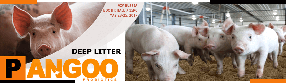
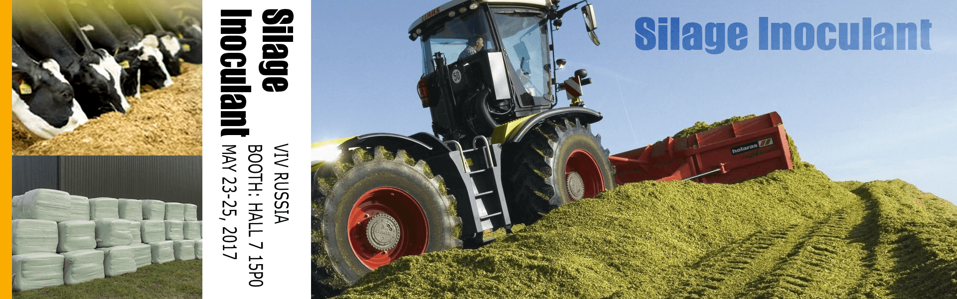
- Deep Litter VS Factory Farming
2017 02/22
- PANGOO will be VIV Russia!
2017 02/23
- HI Antibody level trend after using Avian Influenza Specific Immune Probiotic with avian influenza vaccineHI Antibody level trend after using Avian Influenza Specific Immune Probiotic with avian influenza vaccine HI Antibody level trend after using Avian Influenza Specific Immune Probiotic with avian influenza vaccine (H5 subtype) Weeks 1 2 3 4 5 6 7 8 9 10 11 12 13 14 15 16 H5 Vaccine 0 1.2 1.3 4.2 5.4 6.1 7.3 8.0 7.5 7.0 6.8 6.0 5.6 6.0 5.6 5.5 Immune Probiotics 0 1.2 1.3 4.3 5.5 6.0 7.4 8.2 7.5 7.2 7.0 6.4 5.9 5.8 5.9 5.7 The results showed that the titer of avian influenza antibody increased gradually after immunization with Avian Influenza Specific Immune Probiotic and avian influenza vaccine, and reached a peak after 1 month, and then gradually decreased. Compared with avian influenza vaccine, Avian Influenza Specific Immune Probiotic antibody titer of immune attenuation more slowly. Avian Influenza Specific Immune Probiotic VS avian influenza vaccine (H9 subtype) Measuring with chicken at the age of 14 days, noted the H9 antibody titer; H9 antibody was injected intramuscularly at the age of 15 days; Used Avian Influenza Specific Immune Probiotic at the age of 16 days. Groups 16 days 21 days 30 days Control group 5.16±0.40a 3.12±0.64a 2.87±0.51a Immune Probiotics group 5.00±0.89a 3.37±0.74a 4.80±0.53a Vaccine group 5.00±0.89a 4.47±0.51a 5.25±0.46a Immune Probiotics & Vaccine group 5.00±0.89a 4.55±1.16a 5.65±0.46a The results showed: There were maternal antibodies in egg chickens, and decreased with age. The antibody of Avian Influenza Specific Immune Probiotic AND H9 vaccines both increased, but Avian Influenza Specific Immune Probiotic much faster. Avian Influenza Specific Immune Probiotic combined with H9 vaccine, the effect was significantly better than H9 vaccine group. Antibody titer data for different age groups in broiler flocks (log2) Antibody Groups 2 Weeks 6 Weeks 14 Weeks 26 Weeks 28 Weeks H5 Antibody titer Bivalent Vaccine 0.4 4.6 5.7 6.7 7 Bivalent Vaccine & Immune Probiotics 0.4 5 5.8 7 7.5 H9 Antibody titer Bivalent Vaccine 0.7 4.7 6 6.8 9 Bivalent Vaccine & Immune Probiotics 0.4 5.5 6.3 7 9.3 The antibody titers of H5 and H9 in broiler chickens were increased gradually after vaccination with H5 + H9 bivalent vaccine. The antibody titers of H5 and H9 in broiler chickens were keeping above 51og2 from the 6th weeks. Compared with using of double-seedling only, more immune protection.
2016 12/09
- Newcastle disease Specific Immune Probiotic and Newcastle disease Vaccine used in young Laying hensTest / Feeding data about Newcastle disease Specific Immune Probiotic and Newcastle disease Vaccine used in young Laying hens Test time: 30 days Age of the young laying hens: 1 day old Groups: Control group, Immune Probiotics group, Vaccine group, and Immune Probiotics & Vaccine group. Attention: At the age of 7 days, Immunization by drinking except control group; And at the age of 21 days, Immunization by drinking again only for Vaccine group, and Immune Probiotics & Vaccine group. Antibody titer of Newcastle disease data (log2) Groups 14 days 21 days 30 days Control group 4.16±0.75a 2.50±0.53a 2.25±0.46a Immune Probiotics group 3.90±0.54a 3.65±0.46b 4.37±0.51b Vaccine group 3.83±0.75a 3.25±0.46b 3.80±0.53b Immune Probiotics & Vaccine group 3.83±0.75a 4.67±0.74c 5.40±0.75c There are different effection in Each group, the antibody titer level increased and keep longer time. The result showed: There are maternal antibodies in young laying hens and the maternal antibodies of control group decreased with age. At the age of 21 days and 30 days old, the antibody titer of control group decreased to 2.50 and 2.25, thus, it lost protection ability. The effection of the groups with Immune Probiotics used are better than other groups. Antibody titer of Newcastle disease data (log2) Groups 14 days 21 days 30 days Control group 1985.83 2026.15 2257.72 Immune Probiotics group 2263.12 1808.26 3055.90 Vaccine group 2338.00 1969.55 3692.27 Immune Probiotics & Vaccine group 2458.38 2092.91 3939.14 The result showed: Newcastle disease Specific Immune Probiotics can increase immune level of intestinal mucosal . Because that sIgA content in manure and intestinal of the groups with Immune Probiotics used were effectively increased.
2016 12/09
- Newcastle disease Specific Immune Probiotics and Vaccine used in Laying hensTest / Feeding data about Newcastle disease Specific Immune Probiotic and Newcastle disease Vaccine used in Laying hens Laying hens test base: Our company laying hens farm Hailan brown laying hens at the age of 26 weeks Test time: 33 dats from Nov 16, 2015 Groups: Control group, Immune Probiotics group, and Vaccine group Antibody titer of Newcastle disease data (log2) Groups 26 days 28 days 31 days Control group 9.90 9.83 9.25 Immune Probiotics group 9.50 12.00 13.35 Vaccine group 10.00 11.17 13.07 The result showed that the antiboday titer of both Immune Probiotics group, and Vaccine group increased, but Immune Probiotics group is better and Vaccine group.
2016 12/09
- Newcastle disease Specific Immune Probiotic and Newcastle disease Vaccine Test Data used in BrolierTest / Feeding data about Newcastle disease Specific Immune Probiotic and Newcastle disease Vaccine used in Brolier Feeding test for Broliers at 1-37 days old in 4 groups which include: Control group, Immune Probiotics group(5 billion/brolier), Vaccine group, and Immune Probiotics & Vaccine group. Attention: Newcastle disease vaccine was immunized with eye and drinking water. Antibody titer of Newcastle disease data (log2) Groups 16 days 23 days 30 days 37 days Control group 3.07 2.94 2.88 2.53 Immune Probiotics group 4.17 4.50 5.10 4.80 Vaccine group 3.50 4.30 4.50 4.00 Immune Probiotics & Vaccine group 3.83 4.83 5.17 5.50 The result showed: Both effect of Immune Probiotics group and Immune Probiotics & Vaccine group are better than Vaccine group, and antibody titer higher than control group 1.5-3. The sIgA content in manure of each test group (μg/L) Groups 16 days 30 days Control group 351.12±20.97a 1042.89±211.32aAB Immune Probiotics group 460.06±23.28a 1984.68±262.28aAB Vaccine group 327.12±12.30aA 1574.27±170.46bB Immune Probiotics & Vaccine group 433.62±34.49bB 1780.72±291.92bcC The result showed: At the age of 30 days, the sIgA content in manure of Immune Probiotics group and Immune Probiotics & Vaccine group are much higher than other groups.
2016 12/09
- Data about Escherichia coli Specific Immune Probiotic used in BrolierTest: Feeding data about Escherichia coli Specific Immune Probiotic used in Brolier Test Product: Escherichia coli Specific Immune Probiotic Test for: 200 AA Brolier at the age of 1 day old Test time: for 42 days Test Groups: Blank control group, Natural immunity control group, Immune probiotic test group 1(1 bottle for 2000 brolier), Immune probiotic test group 2(1 bottle for 1500 brolier), Immune probiotic test group 3(1 bottle for 1000 brolier). Test operation: 1. On the 8,9,10,24,25,26 days, using Escherichia coli Specific Immune Probiotic for the 3 immune probiotic test group according the dosage above. 2. Mixed infection with Escherichia coli O1,O35,O2,O78,O26 for the broliers except blank control group, dosage is 1 *108cfu/brolier. Analysis for: Growth performance (body weight), immune organ index, intestinal flora count, E. coli specific antibodies, IgG, sIgA, and the protection of the effect of protection. The Number of Enterobacter Lactobacillus and Escherichia coli in Broilers(*108) The result showed: Escherichia coli Specific Immune Probiotic kept Intestinal microecological balance, powerful protect the chicken from virus. Death Rate & Infection Rate After Mixed infection of Escherichia coli The result showed: Escherichia coli Specific Immune Probiotic can decrease Death Rate & Infection Rate 50-75%. Specific antibody level of Pathogenicity Escherichia coli The result showed: Specific antibody level of Pathogenicity Escherichia coli in the 3 Immune probiotics test group is higher than Blank control group obviously. Data of Broiler body weight, Bursa of fabricius index, and Spleen index The result showed: Broiler body weight, Bursa of fabricius index, and Spleen index in the 3 Immune probiotic test group is higher than Blank control group obviously. Escherichia coli Specific Immune Probiotic on the immune globulin IgA in broilers The result showed: Escherichia coli Specific Immune Probiotic increased the immune globulin IgA in broilers about 20%. Escherichia coli Specific Immune Probiotic on the immune globulin sIgA in broilers The result showed: Escherichia coli Specific Immune Probiotic increased the immune globulin sIgA in broilers about 50-80%. It is proved that Escherichia coli Specific Immune Probiotic has the same effects on Breeding chicken, Laying hens, Meat duck!
2016 12/09
- Data about Salmonella Specific Immune Probiotics used in BrolierTest: Feeding data about Salmonella Specific Immune Probiotic used in Brolier Test product: Salmonella Specific Immune Probiotic Test for: 300 AA brolier at the age of 3 day old Test Groups: Blank control group, Immune probiotics test group, Antibiotic group. Analysis for: Immune index, intestinal flora count, E. coli specific antibodies, IgG, sIgA, and the protection of the effect of protection. Operation: 1. Use anitibiotic for the anitiobic group, and use Salmonella Specific Immune Probiotics for Immune Probiotics group at the aget of 3 days old and 21 days old. 2. Mixed infection with Salmonella at the 30 days old. Data of Specific antibody level and immune globulin at the age of 28 days Salmonella positive rate and Death rate at the 35 days old (After Infection) Immune index, intestinal flora count at the 35 days old (After Infection) The result showed: Immune Probiotics is more effective than antibiotics on treatment of salmonella. Effectively decreased Salmonella positive rate and Death rate, and increased intestinal flora count. At the same time, Salmonella Specific Immune Probiotics decreased the number of Escherichia coli and the number of immune globulin, Bursa of fabricius index, and Spleen index, avoided Secondary infection and mixed infection of Salmonella. The most important is that no drug resistance, because Salmonella Specific Immune Probiotics made by 100% natural probiotics!
2016 12/09
- Data of Salmonella Specific Immune Probiotic used on breeding chickenTest Data of Salmonella Specific Immune Probiotic used on breeding chicken The positive rate of chicken salmonellosis in the breeder farm is high, which can lead to the decline of production performance of adult chicken, decrease of the hatching rate of eggs and cause huge economic loss to chicken industry. For controlling the Salmonella disease, the farmers usually use antibiotics regularly, So that this lead to the drug resistance continued to increase, especially the emergence of multiple drug resistance and drug residues becoming a big problem. For controlling White pullorum well, we applied our Salmonella Specific Immune Probiotic for two big Breeder farms and two big Brolier farms, and controlled the process as : 1. Dinking the Salmonella Specific Immune Probiotic with water at 5 different period which are: Brooding period, incubation period, Pre - production, Pre - fertility peak, after - fertility peak. 2. At the same time, we strictly control the process of regular quarantine, regular immunization, Conventional disinfection, feed purification.... Data of Salmonella positive rate before and after using Salmonella Specific Immune Probiotic
2016 12/09
- Data of Salmonella Specific Immune Probiotic used in Duck paratyphoidTest Data of Salmonella Specific Immune Probiotic used in Duck paratyphoid Test for: 60 Meat duck Test Groups: Blank control group, Immune probiotics test group, Antibiotic group. Analysis for: Immune index, intestinal flora count, E. coli specific antibodies, IgG, sIgA, and the protection of the effect of protection. Operation: 1. Infection with Salmonella (4* 109 cfu/ml) 0.2 ml at the 7 days old. 2. Treatment within 12 hours saperately by antibiotic and immune probiotic. Death Number on the 1st-6th days Control group total death number in the 6 days: 20 Immune Probiotics group total death number in the 6 days: 5 Antibiotics group total death number in the 6 days: 8 Intestinal flora count at the age of 14 days
2016 12/09
- A big difference in EuroTier Hanover, GermanyWhat is the big difference in EuroTier Hanover? During the days in EuroTier Hanover, We have to see Natural feed products accounted for more than 90% of feed products in this exhibition , such as Enzymes, probiotics, or perfect feed formulation product specialized for a certaion animal. Antibiotic products and traditional feed additive companies accounted for a very small proportion. The large feed additive plants only in a small booth, but the biological feed Product companies large-scale display of products, and buyers of the concerns are locked in these Natural products. Though in many developed countries, people still use antibiotic products for promote gowth of the farming animals, but they have taken care much on Natural products, and even try to use it. Benefits of Antibiotic use in Animal Feed The benefits of antibiotics in animal feed include increasing efficiency and growth rate, treating clinically sick animals and preventing or reducing the incidence of infectious disease. By far the major use of antibiotics among these, however, is increased efficiency, i.e. a more efficient conversion of feed to animal products, and an improved growth rate. In chicken feed, for example, tetracycline and penicillin show substantial improvement in egg production, feed efficiency and hatchability, but no significant effect on mortality. Chlorotetracycline, oxytetracyclin and penicillin also show an improved growth rate, but little effect on mortality. Antibiotics in animal feed, in general, are used regularly for increased efficiency and growth rate than to combat specific diseases. Risks of Antibiotics in Animal Feed After animals have been fed antibiotics over a period of time, they retain the strains of bacteria which are resistant to antibiotics. These bacteria proliferate in the animal. Through interaction, the resistant bacteria are transmitted to the other animals, thus forming a colonization of antibiotic resistant bacteria. The bacteria flourish in the intestinal flora of the animal, as well as, in the muscle. As a result, the feces of the animal often contain the resistant bacteria. Transfer of the bacteria from animal to human is possible through many practices. The primary exposure of humans to resistant bacteria occurs in farms and slaughterhouses. Humans clean the feces, which contain the bacteria, of the animals on farms. During the cleaning process, humans may get bacteria on their body and hands. If the body or hands are not properly cleaned, the bacteria could be ingested by the person. Likewise, in slaughterhouses, during slaughter, the intestine is severed. Resistant bacteria are exposed to slaughterhouse workers, which could get the bacteria on their bodies and hands. Transmission occurs when the bacteria is ingested. Along with the previous sources of contamination, humans can get infected by eating meat from animals with resistant bacteria. Even though cooking reduces the survival of the bacteria, some may still survive and infect the human. For example, 1983, 18 people in four midwestern states developed multi-drug resistant Salmonella food poisoning after eating beef from cows fed antibiotics (1). After initial transmission and infection to humans, the transmission to other humans has a couple paths. Transmission can take place through the many mediums (aerosol, physical contact, and bodily fluids) of human contact in the community. An infected individual may also be admitted to a hospital for treatment. Treatment may not work in drug resistant bacteria, therefore, identifying a drug resistant infection. Bacteria is transmitted to other patients via the hospital environment or health care worker=s hands. After transmission, the bacteria will colonize in several of the patients. Colonization in other patients with other resistant bacteria can produce bacteria with multi-drug resistance. Once the patients recover, they are discharged into the community. These patients could potentially infect several community members. Multiple infection could potentially produce a supergerm which is resistant to many drugs due to resistance sharing between bacteria. Use in different livestock In swine production The use of antibiotics to increase the growth of pigs is most studied of all livestock. This use for growth rather than disease prevention is referred to as subtherapeutic antibiotic use. Studies have shown that administering low doses of antibiotics in livestock feed improves growth rate, reduces mortality and morbidity, and improves reproductive performance. It is estimated that over one-half of the antibiotics produced and sold in the United States is used as a feed additive. Although it is still not completely understood why and how antibiotics increase the growth rate of pigs, possibilities include metabolic effects, disease control effects, and nutritional effects.While subtherapeutic use has many benefits for raising swine, there is growing concern that this practice leads to increased antibiotic resistance in bacteria. Antibiotic resistance occurs when bacteria are resistant to one or more microbial agents that are usually used to treat infection. There are three stages in the possible emergence and continuation of antibiotic resistance: genetic change, antibiotic selection, and spread of antibiotic resistance. In production of other livestock The use of drugs in food animals is regulated in nearly all countries. Historically, this has been to prevent alteration or contamination of meat, milk, eggs and other products with toxins that are harmful to humans. Treating a sick animal with drugs may lead to some of those drugs remaining in the animal when it is slaughtered or milked. Scientific experiments provide data that shows how long a drug is present in the body of an animal and what the animal's body does to the drug. Of particular concern are drugs that may be passed into milk or eggs. By the use of 'drug withdrawal periods' before slaughter or the use of milk or eggs from treated animals, veterinarians and animal owners ensure that the meat, milk and eggs is free of contamination. These restrictions include not only poisons or drugs (such as penicillin) which may result in allergic reactions but also contaminants which may cause cancer. It is illegal in the USA to administer drugs or feed substances to animals if they have been shown to cause cancer. One of the main restrictions is the amount that is administered to animals in the industry. These drugs should be administered to healthy livestock at a low concentration of 200 g per ton of feed. The amount distributed is also altered throughout the lifespan of livestock in order to meet specific growth needs. Legality of the use of specific drugs in animal medicine varies according to location. Just as in human medicine, some drugs are available over the counter and others are restricted to use only on the prescription of a veterinary physician. In the USA, theFood and Drug Administration (FDA) requires specific labels on all drugs, giving directions on the use of the drug. For animals, this includes the species, dose, reason for giving the drug (indication) and the required withdrawal period, if any. Federal law requires laypersons to use drugs only in the manner listed. Veterinarians who have examined an animal or a herd of animals may issue a replacement label, giving new directions, based on their medical knowledge. It is illegal in the USA for any layperson to administer any drug to a food animal in a way not specific to the drug label. Over-the-counter drugs which may be used by laypersons include anti-parasite drugs (including fly sprays) and antimicrobials. These drugs can be applied as sprays, creams, injections, oral pills or fluids, or as a feed additive, depending on the drug and the label. In December 2013, the FDA updated its regulations to try to begin reducing use of antibiotics for growth enhancement. Significant lobbying comes from all directions, from those against tighter regulation to those who complain it doesn't go far enough. Currently few policies, regulations and laws exist that promote limitation of antibiotic use on factory farms. In addition, few policies are being created that call for this decrease in antibiotic use. However, numerous state senators and members of congress showed support for the Preventing Antibiotic Resistance Act of 2015 (PARA) and the Preservation of Antibiotics for Medical Treatment Act of 2013 (PAMTA). These acts proposed amendments be made to the Federal Food, Drug and Cosmetic Act which would limit and preserve the use of antibiotics for medically necessary situations. Both of these bills died in Congress in 2015. Use by country European Union The European Union (EU) in 1999 implemented an antibiotic resistance monitoring program and phase out plan for all antibiotic use by 2006. Although the European Union banned the use of antibiotics as growth agents from 2006, its use has not changed much until recently[citation needed]. In Germany, 1,734 tons of antimicrobial agents were used for animals in 2011 compared with 800 tons for humans. On the other hand, Sweden banned their use in 1986 and Denmark started cutting down drastically in 1994, so that its use is now 60% less. In the Netherlands, the use of antibiotics to treat diseases increased after the ban on its use for growth purposes in 2006.In 2011, the EU voted to ban the prophylactic use of antibiotics, alarmed at signs that the overuse of antibiotics is blunting their use for humans. United States In 2011, a total of 13.6 million kilograms of antimicrobials were sold for use in food-producing animals in the United States,which represents 80% of all antibiotics sold or distributed in the United States.Of the antibiotics given to animals fom 2009 through 2013, just above 60% distributed for food animal use are "medically-important" drugs, that are also used in humans.The rest are drug classes like ionophores which are not used in human medicine.Due to concerns about the overuse of antibiotics in food-producing animals, the U.S. Food & Drug Administration has implemented new industry guidelines that will restrict the use of medically-important drugs to uses "that are considered necessary for assuring animal health" and will require veterinary oversight. The food animal and veterinary pharmaceutical industries will need to phase out medically important antimicrobial use by January 1, 2017. China China produces and consumes the most antibiotics of all countries. Antibiotic use has been measured by checking the water near factory farms in China. Measurements have also been taken from animal dung. Half of the antibiotics manufactured in China are used in the production of livestock. It was calculated that 38.5 million kg (or 84.9 million lbs) of antibiotics were used in China's swine and poultry production in 2012. India In 2012 India manufactured about a third of the total amount of antibiotics in the world. Brazil Brazil is the world's largest exporter of beef and the government regulates antibiotic use in the cattle production industry. Concerns about antibiotic resistance More recently, there has been increased concern about the use of anti-microbials in animals (including pets, livestock, and companion animals) contributing to the rise in antibiotic resistant infections in humans. The use of antimicrobials has been linked to the rise of resistance in every drug and species where it has been studied, including humans and livestock. However, the role of antibiotic use in food animals – in contrast to the use of antibiotics in humans – in the rise of resistant infections in humans is in dispute. The use of antimicrobials in various forms is widespread throughout animal industry, and is presented as key to preventing animal suffering and economic loss. It is linked by some activist groups to animal welfare concern, large scale commercial agriculture, international food trade, agricultural protectionist laws, environmental protection (including climate change) and other topics, which make the aims of some groups on both sides of the debate difficult to untangle. Around 70% of all antibiotics administered are used for livestock. Most of the drugs that are given to livestock are misused and incorporated into their diets daily for the purpose of weight gain or to treat illnesses. The overuse of the antibiotic in livestock is harmful to humans because it creates an antibiotic resistant bacteria that can be transferred through several different ways such as: raw meats, consumption of meats, or it can also be airborne. Waste from food-producing animals can also contain antibiotic-resistant bacteria and is sometimes stored in lagoons. This waste is often sprayed as fertilizer and can thus contaminate crops and water with the antibiotic-resistant bacteria. Antibiotic resistance is harmful to humans because it makes them resistant to certain types of drugs for different diseases, and makes it harder for them to fight off infections. The World Health Organization has published a list of Critically Important Antimicrobials for Human Medicine with the intent that it be used "as a reference to help formulate and prioritize risk assessment and risk management strategies for containing antimicrobial resistance due to human and non-human antimicrobial use." Certain antibiotics, when given in low, sub-therapeutic doses, are known to improve feed conversion efficiency (more output, such as muscle or milk, for a given amount of feed) and/or may promote greater growth, most likely by affecting gut flora. Antibiotic Growth Promoters used in Livestock Production drug class effect Bambermycin increase feed conversion ratio and weight gain in chickens, beef cattle, swine, and turkeys. Lasalocid Ionophore increase feed conversion ratio and weight gain in beef cattle. Monensin Ionophore increase feed conversion ratio and weight gain in beef cattle and sheep; promotes proficient milk production in dairy cows. Salinomycin Ionophore increase feed conversion ratio and weight gain. Virginiamycin peptide increase feed conversion ratio and weight gain in chickens, swine, turkeys, and beef cattle. Bacitracin peptide increase weight gain and feed conversion ratio in chickens, turkeys, beef cattle, and swine; promotes egg production in chickens. Carbadox increase feed conversion ratio and weight gain in swine. Laidlomycin increase feed conversion ratio and weight gain in beef cattle. Lincomycin increase feed conversion ratio and weight gain in chickens and swine. Neomycin/ oxytetracycline increase weight gain and feed conversion ratio in chickens, turkeys, swine, and beef cattle Penicillin increase feed conversion ratio and weight gain in chickens, turkeys, and swine. Roxarsone increase feed conversion ratio and weight gain in chickens and turkeys. Tylosin increase feed conversion ratio and weight gain in chickens and swine. Research into alternatives Increasing concern due to the emergence of antibiotic resistant bacteria has led researchers to look for alternatives to using antibiotics in livestock. Probiotics, cultures of a single bacteria strain or mixture of different strains, are being studied in livestock as a production enhancer. Prebiotics are non-digestible carbohydrates. The carbohydrates are mainly made up of oligosaccharides which are short chains of monosaccharides. The two most commonly studied prebiotics are fructooligosaccharides (FOS) and mannanoligosaccharides (MOS). FOS has been studied for use in chicken feed. MOS works as a competitive binding site, as bacteria bind to it rather than the intestine and are carried out. Bacteriophages are able to infect most bacteria and are easily found in most environments colonized by bacteria, and have been studied as well. In another study it was found that using probiotics, competitive exclusion, enzymes, immunomodulators and organic acids prevents the spread of bacteria and can all be used in place of antibiotics. Another research team was able to use bacteriocins, antimicrobial peptides and bacteriophages in the control of bacterial infections. While further research is needed in this field, alternative methods have been identified in effectively controlling bacterial infections in animals. All of the alternative methods listed pose no known threat to human health and all can lead the elimination of antibiotics in factory farms. With further research it is highly likely that a cost effective and health effective alternative could and will be found.
2016 11/25
- Lysine MSDSMaterial Safety Data Sheet (MSDS For L-Lysine HCL) 1.Product- and Company information Product Name: L-Lysine HCL 98.5% feed grade Chemical Name: 2.6 Diaminohexanoic Acid Synonyms:Lysine Hydrochloride *L-Lysine Hydrochloride *Lysine Monohydrochloride *L-Lysine Monohydrochloride Chemical family: Amino acid Composition: As Lysine 78.8% Formula: C6H14N2O2HCl MW: 182.65 CAS No.: 657-27-2 2.Composition / Information On Ingredients Chemical Name CAS No % EINECS# L-(+)-Lysine Monohydrochloride 657-27-2 100% 211-519-9 3.Hazards Identification This product is neither toxic nor dangerous for health. 4. First Aid Measures In case of eye- or skim irritation rinse with water, if the irritation Continues consult a doctor. 5. Fire Fighting Measures Extinguishing media: water, foam, CCh Fire danger: None 6. Accidental Release Measures: Clean up spilled product and keep in resealable, labelled packaging, rinse place out with water. 7.Handling and Storage No specific measures required. Good ventilation recommended 8.Exposure Controls / Personal Protection Inhalation: Dust mask. Hands: Gloves. Eyes: Goggles. 9.Physical And Chemical Properties Form: light beige granules. Smell: odourless Boiling point:not measured Flame point: not applicable Melting point: 260 °C Soluble in water. Relative density:1.28 (water = 1) 10.Stability And Spontaneous Activity Stability:Stable if stored in dry conditions in sealed or closed containers Conditions to Avoid: None Hazardous Decomposition Products:When heated to decomposition, chlorous and Nitrous fumes will be emitted 11. Disposal Considerations Suitable for fill ground, waste disposal following the local regulations and legislation (obligation to inform authorities of the composition). 12.Ecological Information No information available. 13.Toxicological Information RTECS#: CAS# 657-27-2: OL5650000 LD50/LC50: CAS# 657-27-2: Oral, rat: LD50 = 10 gm/kg. Carcinogenicity: L-Lysine hydrochloride -Not listed by ACGIH, IARC, NIOSH, NTP, or OSHA. Epidemiology: No information available. Teratogenicity: No information available. Reproductive Effects: No information available. Neurotoxicity: No information available. Mutagenicity: No information available. Other Studies: No information available. 14.Transport Information The product is NOT classified as dangerous 15. Regulatory Information Classification for water contamination: The product is not listed on the list of products that can be a risk for water. 16. Other Information "IMO" : not dangerous for sea transport ----------------------------------------------------------------------------------------------------- Disclaimer: *************************************************************** Additional references can be taken from the label or the product description. The information given here is correct to the best of our knowledge at the time of writing this sheet. No responsibility can be taken for improper use or handling of the product. *************************************************************** http://pangoogroup.com info@pangoo.biz - 3 -
2016 11/25
- The use of prebiotics and probiotics in pigsThe use of prebiotics and probiotics in pigs Contents 1. The probiotic and prebiotic concept 2. Aspects relevant to the use of probiotics in pigs 2.1 Rearing of pigs 2.2 The porcine digestive tract 2.2.1 Stomach 2.2.2 Small intestine 2.2.3 Large intestine 2.3 Lactic acid bacteria indigenous to pigs 2.4 Detection and identification of lactic acid bacteria in the porcine gastro- intestinal tract 2.5 Immunology 3. Use of prebiotics and probiotics 4. Efficacy and mode of action of probiotics and prebiotics 5. Selection of potential probiotic strains 5.1 The use of gastro-intestinal models to screen cultures for probiotic properties 5.2 The safety of probiotic bacteria 6. Situation in South Africa 7. General discussion 1. The probiotic and prebiotic concept The concept of probiotics evolved at the beginning of the 20th century from a hypothesis first proposed by the Nobel Prize winning Russian scientist Elie Metchnikoff. He suggested that the long and healthy life span of Bulgarian peasants was due to the consumption of fermented milk products (Metchnikoff, 1908). During the last few decades, research on probiotics has expanded beyond bacteria isolated from fermented dairy products to normal microbiota of the intestinal tract (Sanders and Huis in`t Veld, 1999). Vanbelle et al. (1990) defined probiotics as natural intestinal bacteria that, after oral administration in effective doses, are able to colonize the animal digestive tract, thus keeping or increasing the natural flora, preventing colonization of pathogenic organisms and securing optimal utility of the feed. Prebiotics are defined as non-digestible food ingredients that affect the host beneficially by selectively stimulating the growth and/or activity of bacteria in the colon (Gibson and Roberfroid, 1995). This definition was recently amended to `A prebiotic is a selectively fermented ingredient that allows specific changes, both in the composition and/or activity in the gastrointestinal microbiota that confers benefits upon host well-being and health.` In practice, the beneficial bacteria that serve as targets for prebiotics are mostly lactobacilli and bifidobacteria (Gibson et al., 1999; Bouhnik et al., 2004). Unlike probiotics were allochthonous microorganisms are introduced in the gut, and have to compete against established colonic communities, an advantage of using prebiotics to modify gut function is that the target bacteria are already commensal to the large intestine (Macfarlane et al., 2008). However, if for any reason like disease, ageing, antibiotic or drug therapy, the appropriate health-promoting bacteria are not present in the bowel, prebiotics are not likely to be effective. Combinations of prebiotics and probiotics are referred to as synbiotics. Commercial probiotic products often do not meet expected standards in that the composition and viability of the strains may differ from information on the label (Hamilton-Miller et al., 1999; Hamilton-Miller and Shah et al., 2002; Weese, 2002; Fasoli et al., 2003). Another major issue in relation to the application of probiotics is the poor evidence for efficacy based on clinical trials (Klaenhammer and Kullen, 1999). Three issues interfere with the identification of specific health effects of probiotics (Klaenhammer and Kullen, 1999). Firstly, the complexity and variability of the gastro-intestinal environment in relation to gastro-intestinal diseases make it difficult to determine the effect probiotics have on health and disease. Secondly, confusion as to the identity, viability and properties of probiotics lead to strains being incorrectly identified. Lastly, single probiotic strains induce a multitude of effects among different hosts in a test population. A mono-strain probiotic is defined as containing one strain of a certain species whereas multi-strain probiotics contain more than one strain of the same species or genus. The term multi-species probiotics is used for preparations containing strains that belong to one or preferably more genera (Timmerman et al., 2004). Multi-species preparations have an advantage when compared to mono- and multi-strain probiotics (Timmerman et al., 2004). Multi-species probiotics benefit from a certain amount of synergism due to the combination of characteristics from different species. The concept of probiotics plays an important role in animal health. Pig rearing has become an intensive commercial industry. Economic losses due to decreased health and performance brought about by intensive farming practices focused on increased production and low costs, are very important. Major efforts have been made to find different ways to improve the rearing of pigs. Antibiotics have been used successfully for more than 50 years to enhance growth performance and control the spread of disease (Gustafson and Bowen, 1997). Antibiotic resistance is as ancient as antibiotics, protecting antibiotic producing organisms from their own products (Phillips et al., 2004). Antibiotic resistant variants and species that are inherently resistant can dominate and populate host animals. Increased concern exists about the potential of antibiotics in animal feed and their contribution to the growing list of antibiotic-resistant human pathogens (Corpet, 1996; Williams and Heymann, 1998). Although the use of antibiotics for growth promotion is still allowed in certain countries, including the United States, Australia and South Africa, several European countries have implemented strict legislation to prevent the incorporation of antibiotics in animal feed (Ratcliff, 2000). In 1986 Sweden was one of the first countries to ban the incorporation of low-dose antibiotics into animal feed. The question remains, does the use of antibiotics in production animals pose a risk to human health? In a recent review, it was stated that the actual danger appears small and the low dosages used for growth promotion (generally below 0.2% per ton feed) could not be regarded as a hazard (Phillips et al., 2004). Antibiotics are used in animals and humans, and most of the resistance problem in humans arises from medicinal use. Resistance may develop in bacterial populations present in production animals, and resistant bacteria can contaminate animal-derived food, but adequate cooking destroys most bacteria. Growth-promoting antibiotics predominantly active against Gram-positive bacteria have very little or no effect on the antibiotic resistance of salmonellae and consequently on infections caused by salmonellae. In some parts of the world, antibiotics used to treat animals and added to feed as growth promoters may have adverse effects when associated resistance is taken into account. The same antibiotics are often used to treat humans (Phillips et al., 2004). In contrast, Piddock (2002) could not find clear evidence that antibiotic-resistant bacteria isolated from animals, cause infections in humans, for example quinolone-resistant strains of Salmonella serovar Typhimurium DT104 are not transmitted through production animals. The flouroquinolones used therapeutically in animals appear to pose little threat to human health. Flouroquinolone resistance was recorded in bacteria isolated from humans, in countries where the use of this growth promoter is banned such as Sweden, Finland and Canada (Rautelin et al., 1993; Sjögren et al., 1993; Gaudreau and Gilbert, 1998). Faecal flora isolated from a healthy person may contain antibiotic resistant enterococci, but most enterococci isolated from animals do not colonize the human intestine (Dupont and Steele, 1987; SCAN, 1996, 1998; Bezoen et al., 1999; Butaye et al., 1999; Acar et al., 2000). E. coli resistance is more likely to be driven by antibiotic use in humans, although an animal origin for at least some clinical isolates cannot be excluded (Gulliver et al., 1999). The banning of antibiotic usage in animal feed remains a controversial issue especially in the way that it affects farming with production animals. Many natural substances have been investigated as alternatives to conventional chemotherapeutic agents (Turner et al., 2002). Probiotics are one approach used to improve piglet health and deal with intestinal problems encountered during rearing (Vanbelle et al., 1990). Other approaches include acidification of feed or water (Chapman, 1988), altering dietary formulations for small piglets, the development of feeds with lower protein content (Lawrence, 1983), and vaccination with attenuated pathogens or with strains genetically modified (Greenwood and Tzipori, 1987; Trevallyn-Jones, 1987). The administration of growth hormones, somatostatin immunization and enzyme supplementation were also considered as alternatives to antibiotic treatment (Thacker, 1988). Treatment with psychopharmacological drugs (Björk et al., 1987), utilization of the lacto-peroxidase system (Reiter, 1985) and stimulation of hormone-like proteins (anti-secretory factors) capable of reversing intestinal hyper secretion to reduce symptoms of diarrhoea (Lönnroth et al., 1988) were proposed. Some esoteric substances such as zeolite have reduced diarrhoea in piglets and increased feed efficiency (Mumpton and Fishman, 1977). Natural substances that enhance growth performance and immune function in pigs include plant products such as seaweed, saponins extracted from certain desert plants, spices and herbs (Turner et al., 2002). Probiotic preparations may be incorporated in prophylactic agents and it is important to know the mode of action to anticipate the dosage levels (Jonsson and Conway, 1992). The use of probiotics should not exclude other alternatives and a combination of treatments may be complimentary and more effective. 2. Aspects relevant to the use of probiotics in pigs To understand the effect probiotics have on piglets, a thorough understanding of aspects affecting the rearing of pigs and their digestive tract is needed. 2.1 Rearing of pigs One of the major problems in the rearing of pigs is the high mortality rate (ca. 20%) up to weaning age (Bäckström, 1973). In piggeries pre-weaning mortality is caused by diarrhoea, overlay, splay leg, anaemia, bacterial septicaemia, necrotic enteritis, cold exposure and/or congenital defects (Fahy et al., 1987). Piglets in a piggery are often born immature, which renders them more vulnerable to infections. Neonatal diarrhoea often manifests 48 h after birth and is largely attributed to the enterotoxic E. coli strains K88 (most frequent), K99, 987P or F41 (De Graaf and Mooi, 1986). Salmonella spp., Campylobacter spp., Cryptosporidium, transmissible gastroenteritis virus, rotavirus, porcine adenovirus and coronavirus may also cause diarrhoea (Tzipori, 1985; Fahy et al., 1987). The disease manifests by hypersecretion of fluids across the gut wall and into the lumen, triggering the host`s immune system through the various toxins produced. Piglets are particularly susceptible to diarrhoea during the first three weeks after birth and at weaning age (21- to 28-days-old). During the first days, the piglet is protected by maternal immunoglobulins in the colostrum (Porter, 1969). Post-weaning diarrhoea occurs approximately 4 to 10 days after weaning. Enteropathogenic E. coli is the major pathogen (Fahy et al., 1987). Many theories have been proposed as to why disease occurs at weaning. One hypothesis is the sudden deprivation of maternal antibodies and other protective factors in the sow`s milk. Another possibility is sudden changes in diet and/or a compromised metabolism (Fahy et al., 1987) that may lead to particles being metabolized by pathogens, which results in an increase of cell numbers. Changes in temperature, humidity and other environmental conditions may also affect the animal`s immune system (Carghill, 1982), leading to diarrhoea (Björk et al., 1984). Traditionally, pigs have been weaned after 7 to 10 weeks, but piglets are now weaned after 3 to 4 weeks. At this young age the intestinal tract is not able to digest the diets developed for older pigs (Cranwell and Moughan, 1989). The correct feed formulation is thus of critical importance. Feed should contain easily digestible components. During the fattening stage (six months and older) swine dysentery is a problem and feed should be adapted to achieve desirable performances. 2.2 The porcine digestive tract The length of the gastro-intestinal tract (GIT) in the newborn pig is only two meters compared to 20 meters in a mature animal (Slade, 2004). Probiotics need to resist low pH and proteolytic enzymes in the digestive tract. The retention time, mixing of the ingested material with gastric juices and previous digesta, influences the survival of the administered strains. In the anterior part of the small intestine, the most important defense is the fast flow rate that prevents microbial overgrowth, provided the microorganisms do not attach to the epithelium. The presence of bile in this region also represses survival and activity of the microorganisms. In the caecum and large intestine probiotics have to compete with a stable indigenous microflora in the healthy host animal, but the passage rate is slower and the microorganisms establish easier (Jonsson and Conway, 1992). 2.2.1 Stomach The entrance of the stomach has the same type of keratinized squamous non-secreting epithelium as the esophagus (Noakes, 1971). In this region epithelial cells are released continuously and are covered with intestinal bacterial cells including lactobacilli (Lipkin, 1987). Released squamous cells colonized by these bacteria may help to regulate the composition of the digestive microflora by ensuring dominance of the lactic acid bacteria (Fuller et al., 1978; Barrow et al., 1980). In the stomach, gastric juices containing mucus, HCl, proteolytic enzymes and low pH are factors influenced by the age of the animal. The stomach pH may be as low as 2.0 in an adult pig, but as high as 5.0 in milk-fed piglets (Slade, 2004). The intestinal pH of pigs at different ages is listed in Table 1. The degree of mixing and the rate at which contents pass through the stomach influence the effectiveness of the digestion process. Mixing of the digesta depends on dry matter content and particle size. Liquid feed and finely ground feed are mixed more easily than drier or coarsely ground cereal diets (Maxwell et al., 1970). Table 1 pH Values in the digestive tract of pigs Age Stomach Small intestine Caecum Colon Anterior Posterior Neonatal 4.0 - 5.9 6.4 – 6.8 6.3 – 6.7 6.7 – 7.7 6.6 – 7.2 Pre-weaned 3.0 – 4.4 6.0 – 6.9 6.0 – 6.8 6.8 – 7.5 6.5 – 7.4 Weaned 2.6 – 4.9 4.7 – 7.3 6.3 – 7.9 6.1 – 7.7 6.6 – 7.7 Adult 2.3 – 4.5 3.5 – 6.5 6.0 – 6.7 5.8 – 6.4 5.8 – 6.8 Compiled from Smith and Jones (1963), Smith (1965), Boucourt and Ly (1975), Clemens et al. (1975), Braude et al. (1976), Cranwell et al. (1976), Barrow et al. (1977), Schulze (1977), Schulze and Bathke (1977). 2.2.2 Small intestine The acidified portions of digesta entering the duodenum are mixed with bile, pancreatic juice, enzymes and other substances. The pH increases in the small intestine, but variations are less than encountered in the stomach. The difference between piglets and adult pigs is less pronounced (Kidder and Manners, 1978). Variations are large in the duodenum (pH 2.0 to 6.0) and progressively smaller towards the ileum (pH 7.0 to 7.5). The activity of microflora in the distal part of the small intestine lowers the pH in this region (Friend et al., 1963). It normally takes 2.5 h for a food particle to pass through the small intestine (Kidder and Manners, 1978). At this flow rate, it is difficult for bacteria to multiply fast enough to prevent being washed out and probiotics should be administered in sufficient dosages. Attachment to epithelial cells is a prerequisite for bacteria to colonize the small intestine. Volumes measured for the small intestine can be as much as 0.1, 0.6 and 20 L for very young, weaned and adult pigs, respectively (Vodovar et al., 1964). 2.2.3 Large intestine The large intestine consists of the caecum, spiral colon and the distal colon. The rate of passage is slower compared to the small intestine, leading to the establishment of a dense and complex anaerobic microflora. The first part of a meal reaches the anus after 10 to 24 h, but the mean retention time is much more variable and can be two to four days (Kidder and Manners, 1978). The large intestine can hold volumes up to 0.04, 1.0 and 25.0 L for very young, weaned and adult pigs, respectively (Kidder and Manners, 1978). The pH of the large intestine remains at approximately 6.0 (Kidder and Manners, 1978). 2.3 Lactic acid bacteria indigenous to pigs The pig is a monogastric animal in which the foregut (stomach and small intestine) is colonized by a relatively large variety of microflora. Bacteria in the small intestine survive low pH conditions better and bacterial numbers are generally high (107 to 109 cfu/ml) in this section of the GIT (Conway, 1989). Lactic acid bacteria (LAB), mostly Lactobacillus and Streptococcus spp. dominate the small intestine (Fuller et al., 1978). LAB in the foregut helps the young pig to decrease the stomach pH by the production of lactic acid and other organic acids, mainly from lactose (Cranwell et al., 1976; Barrow et al., 1977). LAB may regulate the microflora of the small intestine by migrating with the digesta passing down the GIT (Fuller et al., 1978). Gram-negative bacteria dominate the caecum (Robinson et al., 1981) and Gram-positive species the colon (Salinatro et al., 1977). Species often found in the porcine digestive tract are Lactobacillus acidophilus, Lactobacillus delbreuckii, Lactobacillus fermentum, Lactobacillus reuteri, Lactobacillus salivarius, Enterococcus bovis, Enterococcus durans, Enterococcus faecalis, Enterococcus faecium, Streptococcus intestinalis, Streptococcus porcinus, Streptococcus salivarius, Bifidobacterium adolescentis and Bifidobacterium suis (Raibaud et al., 1961; Zani et al., 1974; Barrow et al., 1977; Fuller et al., 1978; Collins et al., 1984; Robinson et al., 1984; Robinson et al., 1988). The selection and establishment of the indigenous LAB in the neonatal pig develops progressively from birth (Sinkovics and Juhasz, 1974; Schulze, 1977). A succession of Lactobacillus spp. occurs in the small intestine (Tannock et al., 1990) L. reuteri colonize animals on the first day of birth, with the L. acidophilus group appearing one week after birth (Naito et al., 1995). Lysozyme in sow`s milk has a significant effect on bacterial colonization of the pre-weaned piglet (Schulze and Müller, 1980). Colostrum from the sow`s milk provides a protective effect against pathogen-induced diarrhoea (Ducluzeau, 1985). Adverse conditions may lead to changes in the intestinal flora. Markedly lower numbers of lactobacilli and bifidobacteria were detected in the foregut of piglets deprived of water and food for 72 h, while numbers of E. coli increased (Morishita and Ogata, 1970). 2.4 Detection and identification of lactic acid bacteria in the porcine gastro-intestinal tract Understanding of the complex natural bacterial communities that colonize the GIT of monogastric mammals such as pigs and humans is far from complete. The identification of faecal flora by time-consuming methods where intestinal bacteria had to be isolated and cultured, revealed considerable species diversity (Moore and Holdeman, 1974; Salinatro et al., 1977; Moore et al., 1987). Over the past decade, molecular methods have been developed that may be used to study the diversity of the gut microflora (Wilson and Blitchington, 1996). Molecular biology plays an important role in the field of probiotics, where it is used as a taxonomic tool. Current techniques like genetic fingerprinting, gene sequencing, oligonucleotide probing and specific primer selection, discriminate closely related bacteria with varying degrees of success (McCartney, 2002). Additional methods that are used include DGGE, temperature gradient gel electrophoresis (TGGE) and fluorescent-in-situ-hybridization (FISH). FISH can be used to great effect in the identification of intestinal microorganisms. By applying fluorescently labeled oligonucleotides, individual whole fixed cells can be identified in situ (Delong et al., 1989; Amann et al., 1990). FISH can be implemented in the detection of probiotic bacteria since rDNA targeted specific oligonucleotide probes can be designed for the probiotic strains administered, that would enable detection of the cells in the mucus. The addition of fermentable carbohydrates supports the growth of lactobacilli in the ileum and colon of weaning piglets. Future molecular biology studies on probiotics and gut flora will lead to a better understanding of the activity and function of microflora (McCartney, 2002). The quest will be to demonstrate the role of probiotic bacteria in vivo. 2.5 Immunology In the healthy adult pig, immunoglobulins are released into the digestive tract and contribute to the host`s defense against infection. This immune defense starts to function soon after birth and continues up to about 3 weeks of age (Jonsson and Conway, 1992). After 3 weeks, IgA is secreted and provides immune protection (Porter, 1969). Sow milk immunoglobulins inhibit the growth of E. coli (Wilson and Svendsen, 1971), adhesion to enterocytes (Nagy et al., 1979) and neutralizes toxins (Brandenburg and Wilson, 1973). At weaning, the piglet is suddenly deprived of milk antibodies and some non-immunological factors such as lactoferrin, transferrin, vitamin B12-binding protein and the bifidus factor (Cranwell and Moughan, 1989). Although the immune system of piglets is fully functional at the time of weaning, it may need to be stimulated to prevent diarrhoea. Probiotics may stimulate the immune system (Perdigón et al., 1987; Shahani et al., 1989). Hypersensitivity responses in the early-weaned piglet may be induced by dietary components. The intake of small amounts of certain proteins before weaning, particularly soy, sensitizes the immune system (Newby et al., 1984). Mild diarrhoea and some intestinal disturbances may result, leading to increased susceptibility to pathogenic infections. 3. Use of probiotics and prebiotics The interest in probiotics increased during the 1940s, followed by a decline. However, interest is escalating again as can be seen from the number of recent publications. Emphasis has shifted from using milk fermented with microbes to selecting for indigenous bacteria. The species used in probiotic products for pigs include L. acidophilus, Lactococcus lactis, L. reuteri, combinations of Lactobacillus spp., E. faecalis, E. faecium, Bacillus licheniformis, Bacillus subtilis, Bacillus subtilis var. toyoi, Bifidobacterium bifidum, Bifidobacterium pseudolongum, Bifidobacterium thermophilus, Clostridium butyricum, Saccharomyces spp. and other yeasts. Mixed combinations of organisms used, include Pediococcus acidilactici, Lactobacillus plantarum, Lactobacillus casei, L. fermentum, Lactobacillus brevis, Lactobacillus delbreuckii subsp. bulgaricus, L. casei, Streptococcus salivarius subsp. thermophilus, L. plantarum, L. acidophilus and E. faecium (Jonsson and Conway, 1992). Lactobacilli are strong acid producers and seldom pathogenic (Sharpe et al., 1973; Sharpe, 1981). Certain strains of E. faecalis and E. faecium are pathogenic (Hardie, 1986; Mundt, 1986). However, some non-pathogenic enterococci are incorporated in probiotic products (Strompfová et al., 2004). Many enterococci produce antimicrobial substances (enterocins) and have an effect on spoilage organisms (Cintas et al., 1997; Sabia et al., 2002). Enterococci can be used as probiotic organisms because of high growth rate, adhesion ability and production of enterocins (Maia et al., 2001). Non-pathogenic strains of certain E. coli can be administered to prevent subsequent colonization of other pathogenic bacteria in the GIT (Duval-Iflah et al., 1983). One of the best examples of probiotic E. coli is strain Nissle 1917 (EcN), serotype O6:K5:H1 (Blum et al., 1995). This strain lacks typical virulence genes and prevents the invasion of Yersinia enterocolitica, Shigella flexneri, Legionella pneumophila and Listeria monocytogenes (Altenhoefer et al., 2004). Probiotic preparations should be administered soon after birth, when disease is anticipated (preventive or curative). Administration could be orally (although this could be very stressful to the animals), or dispensed in water or feed (pelleted or ground). Probiotic bacteria can be given as viable organisms in wet, frozen or freeze-dried preparations or pastes (Tournut, 1989), or as fermented products (Pollman et al., 1984). Pelleting involves high temperatures and pressures that may be lethal to microorganisms (Jonsson and Conway, 1992). Some streptococci and Bacillus spp. are less affected by heat and may survive, but lactobacilli are more sensitive. Growth conditions of the bacteria, harvesting methods and exposure conditions prior to freeze-drying also influence survival of the cells. Normal indigenous microflora of a healthy pig may not establish in the GIT when piglets are moved directly after birth into a scrupulously clean environment, or after antibiotic treatment. Preparations of LAB can be administered at these times to initiate the natural sequential colonization of the digestive tract (Cranwell et al., 1976). With normal pig rearing the piglets stay in close contact with the sow for the first weeks, and will be colonized by LAB. On farms with a high incidence of diarrhoeal disease, it may be appropriate to introduce a probiotic strain as early as possible to colonize the digestive tract with probiotic strains that inhibit pathogens. The characteristics and mechanisms of action of the specific strains used will determine whether a single or continuous dosage is preferable. Therapeutic doses are 109 to 1012 viable organisms per animal per day or 106 to 107 added to feed (Vanbelle et al. 1990). The number of organisms given should be sufficient to elicit a beneficial response in the host, but should not induce digestive disorders (Jonsson and Conway, 1992). The issue whether administered probiotic microorganisms are transient or adhere in the GIT, influences the dosage required. Transient strains need to be administered at higher levels than strains adhering to and multiplying in the GIT (Conway, 1989). Prebiotics are often administered in conjunction with probiotics. The dominant prebiotics used are fructo-oligosaccharides (FOS), oligofructose and inulin trans-galacto-oligosaccharides (TOS), gluco-oligosaccharides, glyco-oligosaccharides, lactulose, lactitol, malto-oligosaccharides, xylo-oligosaccharides, stachyose and raffinose (Monsan and Paul, 1995; Orban et al., 1997; Patterson et al., 1997; Collins and Gibson, 1999; Patterson and Burkholder, 2003). Although mannan oligosaccharides (MOS) have been used as prebiotics, they do not enrich probiotic bacterial populations, but act by binding and removing pathogens from the intestinal tract and by stimulating the immune system (Spring et al., 2000). The oligomers, galacto-oligosaccharides (GOS), soybean oligosaccharides, lactosucrose, isomalto-oligosaccharides and palatinose revealed prebiotic potential (Manning et al., 2004). The vast majority of studies on prebiotics focused on inulin, FOS, GOS and TOS. The latter group of carbohydrates has a history of safe commercial use (Macfarlane et al., 2008). 4. Efficacy and mode of action of probiotics and prebiotics Administration of probiotic products often gives inconclusive or conflicting results in host animals and determination of the mode of action becomes more difficult (Jonsson, 1985; Tuschy, 1986; Conway, 1989). One important factor to consider is that host susceptibility varies from one animal to the other. Evaluations of probiotic use should include the effect on microflora in the digestive tract. Performance and health can be evaluated by growth rates, feed utilization, number of deaths and occurrence of diarrhoea (Jonsson and Conway, 1992). The clinical conditions in which efficiency of probiotics have been reported range from infectious, allergic and inflammatory to neoplastic, suggesting that a single mechanism of action is unlikely (Marteau and Shanahan, 2003). Various hypotheses exist to explain the mode of action of probiotics, but these remain speculatory (Vanbelle et al., 1990). Health promoting advantages of probiotic preparations include production of antimicrobial substances, organic acids, and prevention of adhesion of pathogenic bacteria in the digestive tract. Other possible modes of action include production of metabolites able to neutralize bacterial toxins in situ or inhibition of their production. An increase in feed conversion by secretion of enzymes from the microflora, stimulation of the immune system, and proliferation in the GIT were also suggested as possible modes of action. Sakata et al. (2003) suggested that probiotics modify the metabolism in the microbial ecosystem of the large intestine by increasing the production of short chain fatty acids (SCFA). This leads to an increase in sodium and water absorption and a decrease in colonic activity. The SCFA act as modulators for required functions to ensure a healthy GIT. One study assessed the body weight, weekly feed intake and feacal consistency after probiotic supplementation One advantage recorded for probiotic supplementation in this study included reduction of weaning diarrhea in piglets (Taras et al., 2007). The exact mechanism of action of probiotics remains largely unknown. Probiotics may contribute to host defense by reinforcing non-immunological defenses and stimulating both specific and non-specific host immune responses (Gill, 2003). Little is known about the relative importance of the probiotic-stimulated mechanisms in host protection. Prebiotics have been shown to possess some immunomodulatory properties. To assess the effects of prebiotics such as FOS and GOS on the immune system, a large number of immunological parameters/markers needed to be assessed (Macfarlane et al., 2008). Measurements of these markers had to take into account the fact that they can be affected by gender and age, and that they might vary because of external factors such as stress, smoking and alcohol intake, which necessitates careful selection of control subjects. The gut contains lymphoid tissue that forms a major part of the body`s immune system. Experimental data obtained suggest that immunomodulation can occur through the use of functional foods such as prebiotics (Macfarlane et al., 2008). Prebiotics like raffinose have been shown to reduce allergic reactions in children (Nagura et al., 2002) and results obtained to date with prebiotics in relation to osteoporosis offer some promise (Macfarlane et al., 2008) but studies are limited. Lactulose, FOS and GOS have laxative effects with lactulose well established in the treatment of constipation (De Schryver et al., 2005). Latest studies indicate that there might be potential use of prebiotics on their own, or in combination with probiotics, to immunomodulate the diseases like rheumatoid arthritis and cancer (Macfarlane et al., 2008) but these studies are however in a very early stage. Research data demonstrating the efficacy of prebiotic application in pigs are scarce compared to human studies (Mountzouris et al., 2006). Most prebiotic oligosaccharides incorporated into swine diets at levels ranging from 5 to 40 g/kg have resulted in mixed but generally not significant effects regarding beneficial modulation of microbial populations determined in various intestinal segments and faeces of pigs (Flickinger et al., 2003; Mikkelsen et al., 2003). Prebiotic inclusion levels in feeds at higher levels have resulted in significantly increased levels of bifidobacteria and lactobacilli in the porcine gut, but at the higher cost of nutrient digestibility (Smiricky-Tjardes et al., 2003). However, depression of nutrient digestibility is linked to animal performance and health, therefore careful assessment is required. It was concluded that in terms of microflora metabolic activity, the substantially higher numerical trends seen in TOS and FOS treatments regarding total volatile fatty acid, acetate concentrations and glycolytic activities, it could be postulated that TOS and FOS promoted saccharolytic activities in the pig colon (Mountzouris et al., 2006). Overall, effects of prebiotics on porcine gut health have often been variable and inconsistent. 5. Selection of potential probiotic strains Probiotic strains are selected based on resistance to lytic enzymes in saliva (lysozyme) and digestive enzymes, growth at low pH and bile salts, and their ability to prevent colonization of pathogenic bacteria. Stimulation of the immune system by the probiotic strains is required to increase cell-mediated immune response. Technological resistance and stability at high temperatures during pelleting, spraying etc. will ensure viability of the probiotic strains after dosage. Cell adhesion is one of the selection criteria that remain controversial. This aspect was derived from the concept of virulence factors in pathogenic bacteria. Adherence promotes certain virulence activities like production of toxins (Edwards and Puente, 1998; Klemm and Schembri, 2000). Similar interactions could be beneficial for probiotic organisms such as lactobacilli. Lactobacilli adhere to mucosal surfaces and thereby limit the adherence of pathogenic bacteria (Kotarski et al., 1997; Kirjavainen et al., 1998). Some lactobacilli lack the ability to bind mucus in vitro (Jonsson et al., 2001). Since many of these non-binders were isolated from mucosal surfaces it may be assumed that the growth environment affects the adhesion properties of bacteria (Jonsson et al., 2001). The adhesion property of probiotic LAB is species-specific (Barrow et al., 1980). Host specificity is a desirable property for probiotic bacteria and is one of the selection criteria (Salminen et al., 1988; Saarela et al., 2000). Adhesion of LAB in relation to host specificity in human, canine, possum, bird and fish mucus were investigated in vitro (Rinkinen et al., 2003). Results indicated that the adhesion trait was not host specific but rather characteristic of the species. This suggests that animal models in probiotic adhesion assays may be more applicable to other host species than earlier thought and highlights the fact that the selection criteria for a probiotic may vary according to the application of the probiotic (Rinkinen et al., 2003). Numerous papers have been published on the isolation and selection of potential probiotic strains (Nemcova et al., 1997; Chang et al., 2001; Gusils et al., 2002). Results obtained with in vivo feeding trials were variable because of the complexity of the intestine and variation between individual animals (Simon et al., 2003). Competitive exclusion products containing undefined cultures were effective in pigs (Fedorka-Cray et al., 1999; Genovese et al., 2000), but the possibility that these products may contain pathogens remains (Gillian et al., 2004). Individual probiotic strains need to be identified before inclusion in a probiotic product (Gillian et al., 2004). Selection characteristics for prebiotics differ from those proposed for probiotics. Prebiotics should not be hydrolyzed by digestive enzymes or absorbed by mammalian tissues. Substances used as prebiotics must selectively enrich beneficial bacteria (Simmering and Blaut, 2001). 5.1 The use of gastro-intestinal models to screen cultures for probiotic properties To select suitable probiotics, the strains have to be studied in the environment where they function. The intestinal tract of humans and animals is not readily available for research purposes. This lead to the development of various models simulating the gastro-intestinal tract (Miller and Wolin, 1981; Veilleux and Rowland, 1981; Edwards et al., 1985; Gibson et al., 1988; MacFarlane et al., 1989; Molly et al., 1993; Veenstra et al., 1993). A unique GIT model was developed at the TNO Nutrition and Food Research Organization, based in the Netherlands. It was the first in vitro model that included features like peristaltic movements, physiological transit characteristics, nutrient absorption and water retention (Veenstra et al., 1993). These unique features made the TNO model very expensive to develop and operate. Potential applications included research on the digestibility of carbohydrates and other food ingredients, interactions of fats and proteins, stability of fat and sugar replacers, availability of minerals and survival of bacteria used in fermented foods and probiotics (Veenstra et al., 1993). The TNO model could be used in both animal nutrition and pharmaceutical research. Latest research included studies of the absorption of mycotoxins in the GIT of pigs (Avantaggiato et al., 2004) and mechanistic studies on the intragastric formation of nitrosamines, resulting in valuable information being obtained regarding the human cancer risk from the combined intake of codfish and nitrate-containing vegetables (Krul et al., 2004). These in vitro models proved a popular tool for research concerning bacterial populations in the GIT and probiotic bacteria administered to animals and humans. Advantages in the use of in vitro models compared to in vivo animal trials and experiments include cost-effectiveness, rapid results, reproducibility and most importantly, no ethical constraints (Veenstra et al., 1993). 5.2 The safety of probiotic bacteria Theoretically, probiotic bacteria may be responsible for side effects such as systemic infections, deleterious metabolic activities, excessive immune stimulation in susceptible individuals and gene transfer (Marteau, 2001). However, only a few cases of side effects in humans have been reported (Marteau, 2001). Limited information is available on the adverse effects of probiotics in animals, especially pigs. Future studies may focus on the degradation of the intestinal mucus layer by probiotics. No mucus degradation was observed in experiments with gnotobiotic rats (Ruseler-van Embden et al., 1995). Antibiotic resistance genes, especially those encoded by plasmids, can be transferred between organisms (Marteau, 2001). This raises the question whether resistance genes can be transferred by probiotics to endogenous flora or to pathogenic microorganisms. Risk of gene transfer depends on the genetic material transferred, nature of the donor and recipient strains and on selective pressure. Probiotics currently used have been assessed as safe in fermented foods, but safety evaluation in microbial food supplements remains controversial since legislation differs between countries (Isolauri et al., 2004). The ability of specific probiotic strains to survive gastric conditions and adhere to intestinal mucosa following oral administration may entail the risk of bacterial translocation, bacteraemia and sepsis (Table 2). It was proved that probiotics improve the microflora in the gut and thus the overall health status of the host animal and that probiotics have [GRAS" (generally regarded as safe) status (Anadon et al., 2006). 6. Situation in South Africa Although the use of antibiotics as a growth stimulant in pig rearing has not been banned thus far in South Africa, the legislation might be implemented in the future. Therefore the Livestock Business division of the Agricultural Research Council (ARC) already has a research programme in place called [Alternatives to antibiotics". At the Animal Feed Manufacturers Association (AFMA) forum in 1998, an overview of the mechanisms of and role of prebiotics and probiotics in animal feeds was presented by the ARC. During the AFMA Forum in March 2007, it was reported that legislation banning the use of antimicrobial growth promoters (AGP`s) was implemented on the 1st of January 2006 in the European Union (EU). The question remained, will the same legislation on AGP`s await South Africa in the future? It was reported at the AFMA forum by Maritz (2006) that varying results were obtained in countries where the use AGP`s were banned. In Denmark, the use of antibiotics as therapeutic treatment increased after the banning of AGP`s, reflecting increasing problems with diarrhoea (Maritz, 2006). The same problem was experienced in Sweden and was addressed by changes in farm management, hygiene, sectioning, zinc supplementation of piglet feed and the use of medicated feed in some herds (Wegener, 2005). The cost of production in the pig industry also increased after the banning of AGP`s (Maritz, 2006). The ARC-Irene is highly committed to the task of finding alternatives to antibiotics, and therefore frequently calls on the livestock, feed and pharmaceutical industries to assist us with research efforts in this regard. It is clear that an outright ban of AGP`s in South Africa will not be feasible (also emphasized by above findings from countries in the EU), thereby supporting the approach of the United States. Specific AGP`s linked directly to human medicinal use will have to be phased out. We hypothesize that a holistic approach will be required as a single alternative is unlikely to be the answer, rather a combination of alternatives might provide products and strategies that will enable AGP`s to be phased out. Human health is of prime importance, especially with the high incidence of HIV/AIDS and other eroding conditions in the population, and the threat of antibiotic resistant bacteria to these individuals as well as to the population as a whole.
2016 11/25
- How's Your Soil Texture?CLAY IS MORE PROMISING THAN IT'S CRACKED UP TO BE Before you try to manage the clay soil in your home garden, it helps to have a bit of background in soil mineralogy. The mineral fraction of a given soil consists of sand, silt, and clay particles. Clay particles are the smallest of the three, silt are intermediate in size, and sand, the largest. Clay particles, bound end to end and side to side in extensive planes, are stacked in a sandwichlike matrix and held together by electrochemical forces. This platelike stacking of horizontally arranged clay particles results in a large surface area. Because individual clay particles are negatively charged, they have the capacity to attract and hold onto, or adsorb, positively charged elements (called cations) such as ammonium, potassium, calcium, magnesium, and other trace elements. Clay soils are relatively fertile because of this capacity to adsorb these important plant nutrients. Conversely, the single, uncharged sand particles in sandy soils lack the capacity to adsorb cations and thus they contribute very little to soil fertility. Soil texture is an inherent property that you cannot change. Instead, direct your efforts toward improving soil structure. Soil texture-The textural designation of a soil is determined by its relative portions of sand, silt, and clay particles, and indicates which of the three most influence the soil's properties. Sand, silt, and clay soil properties are obviously dominated by those respective fractions. For example, clay soils (generally more than 40 percent clay) are often poorly drained. On the other hand, well-drained loam soils are mixtures of sand, silt, and clay in roughly equal proportions, and are well drained. A sandy loam, however, has much more sand and much less clay than does a clay loam. Soil structure-Soil texture is an inherent soil property that you as a gardener cannot change (except through extreme interventions). Instead, you should direct your efforts toward improving soil structure. Soil structure is defined by the manner in which soil particles are assembled as aggregates. In clay soils, clay particles are typically arranged along a horizontal plane in a platelike structure. When these horizontal aggregations are stacked high and consolidated over time, they can be quite tight and sticky. Your aim in improving soil structure is to achieve a looser, more crumbly or granular structural aggregation. A soil with the latter structure has a friable consistency and good tilth. Tilth-Tilth is the physical condition of the soil as it relates to ease of tillage, seedbed quality, ease of seedling emergence, and deep root penetration. A soil that drains well (yet has water-holding capacity), does not crust, takes in water rapidly, facilitates aeration, and does not make clods is said to have good tilth. And with the right management strategy, good tilth is achievable in a clay soil. CLAY DEMANDS SHORT-TERM AND LONG-RANGE STRATEGIES My initial management strategy centered on adding as much organic matter as deeply as I could, because, generally, the application of organic matter to soil improves both structure and tilth, and contributes to improvements in overall soil health. My long-range management strategy attempts to build available organic carbon and humus and to promote nutrient cycling through regular applications of compost, manure, and other organic matter, the incorporation of cover crops as green manures, rotations that include grasses and legumes, and reduced tillage. Organic matter builds soil tilth in a couple of ways. First, the organic matter coats soil particles, physically separating clay particles and aggregates from each other. Second, and more important, microorganisms that degrade organic matter produce byproducts called glomalin that bind individual clay particles together into aggregates. Particle aggregation in the topsoil reduces crusting, increases the rate of water infiltration, and reduces erosion and runoff. Though my ultimate goal continues to be to increase soil organic matter content in the garden, I was less concerned initially with the numbers than I was with achieving the benefits described above. I understood that in southeastern soils, it would be very difficult to increase the percentage of organic matter in the soil very quickly. For one thing, even large amounts of organic matter initially incorporated in the soil will rapidly break down. The organic matter fraction remaining may resist further degradation for years or even decades, but stable increases in this fraction, called the humic fraction or humus, occur very slowly. Thus, any increase in stable organic matter is necessarily a long-term goal. COMPOST PLAYS A KEY ROLE Composts are integral to my clay soil management plan. Because of the humified nature of compost and its low concentrations of oxidizable carbon and available nitrogen, compost is relatively resistant to further decomposition, and additions of compost to the soil over time can increase the soil's organic carbon and humic matter content. I add compost not so much to provide nutrients as to provide stabilized organic matter that will improve the physical properties of the soil. Read articles about composting ... I'm fortunate to have a commercial compost supplier close by my home, so I can take advantage of very reasonable bulk pricing, along with a guaranteed chemical analysis that allows me to apply the compost at appropriate rates. There is absolutely no way I could have built a garden in my clay soil without the 16-cubic-yard tandem truckloads of compost that rolled onto my property. Moving that compost one wheelbarrow load at a time to my vegetable and flower beds is a weekend ritual. The incorporation of the compost into the soil has been more problematic. During the initial, pre-garden phase of my soil improvement plan, I managed to break up 10 inches of hard-packed clay with endless swings of my trusty mattock. I spread compost over the surface of the beds an inch at a time and rototilled it in. I was careful not to work the clay when it was too wet, because clay worked wet can result in some tenacious clods, very reluctant in their willingness to ever come apart again. I tried to work the compost deep into the soil, and along with it, I added lime and phosphorus, the clay soil in my garden needing both. When surface-applied, neither of these materials moves down through the soil, so incorporation to ample depth is very important to permit roots to grow into the subsoil. After 3 to 4 inches of compost, I had the "raised bed" I wanted. I called it quits (for the time being) and planted my garden. In the years since, I have been back through most of the beds several times with the mattock and more compost, trying to get organic matter and lime incorporated deeply to break up the "seal" that forms between amended and unamended soil, and to get my beds raised back up. It's readily apparent that organic matter decomposes very quickly in the vegetable beds. In spite of all the amendments, the beds no longer seem "raised," and there's not much visible evidence of all the compost I've added. However, I can tell by the color, now a dark brown instead of red, and by the aggregation of the soil particles into a crumbly structure (of a kin with coffee grounds) that humus is accumulating. Reworking a bed is always a fall practice when my rotation calls for fall-planted garlic or when potatoes or root crops will be planted early the following spring. I just can't count on the beds being dry enough in spring to work the clay deeply without messing up the structure I've worked so hard to build. COVER CROPS KEEP ON GIVING Along with adding compost, a second strategy for adding organic matter to my clay soil is cover cropping. I plant cover crops any time the beds in my garden would otherwise be unplanted or fallow. Fallow periods provide little additional organic biomass while allowing the decomposition of organic matter in the soil to continue. Cover crops provide me with a variety of services. They contribute to the improvements in soil structure that I described earlier. They reduce erosion and increase infiltration. And they can smother weeds and even suppress weed seed germination. Many cover crops will also suppress pathogenic nematodes, for example root knot nematode. I take a machete and scalp the cover crop at the surface, killing it but leaving the biomass in place. Leguminous cover crops can fix significant amounts of nitrogen for use by subsequent crops. Through symbiotic associations with legumes, Rhizobia bacteria convert atmospheric nitrogen into an organic form that legumes use for growth. How much fixation depends on the length of the growing season, the local climate, and soil conditions. Nonleguminous cover crops, principally grasses or small grains, do not fix atmospheric nitrogen, but can be effective in recycling residual soil nitrogen remaining in the root zone at season's end, reducing leaching losses from the soil during the winter months. I often plant mixtures of grasses and legumes in the fall to reap the benefits of both. This past fall I planted hairy vetch and cereal rye, a proven mixture here. Crimson clover and rye or oats also do well in my area. In mid-May, I become a terminator, literally putting these annual plants down to prepare the beds for summer vegetables. By then, the winter cover crops have provided my annual organic matter addition, as well as enough nitrogen fertilizer to grow my summer crops. THE LESS YOU DISTURB AMENDED CLAY THE BETTER Because of the many benefits that accrue to soil quality, it is clearly in your short-term interest to incorporate organic matter regularly and deeply, at least when beginning the process of whipping a clay soil into shape. Soil particle aggregation and aggregate stability, water holding capacity, drainage, nutrient retention, and plant root growth are all increased when organic matter is incorporated. However, major losses in soil organic matter content can take place when the soil is inverted or mixed annually by tillage. Extensive tillage stimulates microbial activity (gives the little guys an appetite, so to speak), and the consumption of mass quantities of organic matter ensues. After your clay soil becomes more friable and you have provided a deep root zone for your garden plants, you should consider reducing tillage. Switching to a minimum-till system increases soil organic matter, soil organic carbon, total nitrogen, and soil microbial biomass carbon and nitrogen content, especially at the surface and in the top 2 to 4 inches. Because the soil remains undisturbed, fertilizers and other soil amendments do not become homogenized in the tillage layer. Plant feeder roots, therefore, tend to proliferate in the top 2 inches of fertile topsoil. For a number of years now, I have been experimenting with no-till vegetable planting in the spring. I take a big knife (i.e., a machete), make sure there are bandages available nearby, and scalp the cover crop at the surface, killing it but leaving the biomass in place. I pull the residue apart just enough to dig a small hole with a trowel, drop in my tomato, pepper, or melon transplant, and pull the mulch back around the stem. I plant large seeds the same way, except that I leave a mulch-free window for sunshine to strike the seedling when it emerges. For small-seeded vegetables, I generally stir the soil a bit more and leave a strip of mulch-free surface above the seed. Fruiting is delayed a bit because the soil is cooler for a time, but overall production is not sacrificed. The mulch reduces weed growth and prevents soil that may carry overwintering disease pathogens from splashing up on foliage. Infiltration of irrigation water and rainfall improves, as does soil moisture retention. The residue decomposes much more slowly than if I tilled it in. Slower decomposition can result in more favorable timing of the release of nitrogen from the legume cover crop.
2016 10/28
- Classification of Photosynthetic BacteriaOxygenic photosynthetic bacteria perform photosynthesis in a similar manner to plants. They contain light-harvesting pigments, absorb carbon dioxide, and release oxygen. Cyanobacteria or Cyanophyta are the only form of oxygenic photosynthetic bacteria known to date. There are, however, several species of Cyanobacteria. They are often blue-green in color and are thought to have contributed to the biodiversity on Earth by helping to convert the Earth`s early oxygen-deficient atmosphere to an oxygen-rich environment. This transformation meant that most anaerobic organisms that thrived in the absence of oxygen eventually became extinct and new organisms that were dependent on oxygen began to emerge. Cyanobacteria are mostly found in water but can survive on land, in rocks, and even in animal shells (or fur), and in coral. They are also known to be endosymbiont, which means they can live within the cells or body of another organism in a mutually beneficial way. Cyanobacteria also tend to live in extreme weather conditions, such as Antarctica, and are interesting to scientists because they may indicate a chance for life on other planets such as Mars. Anoxygenic photosynthetic bacteria consume carbon dioxide but do not release oxygen. These include Green and Purple bacteria as well as Filamentous Anoxygenic Phototrophs (FAPs), Phototrophic Acidobacteria, and Phototrophic Heliobacteria. Let`s look at the differences between these types of bacteria a little more closely. Purple bacteria can be divided into two main types – the Chromatiaceae, which produce sulfur particles inside their cells, and the Ectothiorhodospiraceae, which produce sulphur particles outside their cells. They cannot photosynthesize in places that have an abundance of oxygen, so they are typically found in either stagnant water or hot sulfuric springs. Instead of using water to photosynthesize, like plants and cyanobacteria, purple sulfur bacteria use hydrogen sulfide as their reducing agent, which is why they give off sulfur rather than oxygen. Purple bacteria are probably the most widely studied photosynthetic bacteria, being used for all sorts of scientific endeavors including theories on possible microbiological life on other planets. Purple non-sulfur bacteria do not release sulfur because instead of using hydrogen sulfide as its reducing agent, they use hydrogen. While these bacteria can tolerate small amounts of sulfur, they tolerate much less than purple or green sulfur bacteria, and too much hydrogen sulfide is toxic to them. Green sulfur bacteria generally do not move (non-motile), and can come in multiple shapes such as spheres, rods, and spirals. These bacteria have been found deep in the ocean near a black smoker in Mexico, where they survived off the light of a thermal vent. They have also been found underwater near Indonesia. These bacteria can survive in extreme conditions, like the other types of photosynthetic bacteria, suggesting an evolutionary potential for life in places otherwise thought uninhabitable. Phototrophic Acidobacteria are found in a lot of soils and are fairly diverse. Some are acidophilic meaning they thrive under very acidic conditions. However, not much is known about this grouping of bacteria, because they are fairly new, the first being found in 1991. Phototrophic Heliobacteria are also found in soils, especially water-saturated fields, like rice paddies. They use a particular type of bacteriochlorophyll, labelled g, which differentiates them from other types of photosynthetic bacteria. They are photoheterotroph, which means that they cannot use carbon dioxide as their primary source of carbon. Green and red filamentous anoxygenic phototrophs (FAPs) were previously called green non-sulfur bacteria, until it was discovered that they could also use sulfur components to work through their processes. This type of bacteria uses filaments to move around. The color depends on the type of bacteriochlorophyll the particular organism uses. What is also unique about this form of bacteria is that it can either be photoautotrophic, meaning they create their own energy through the sun`s energy; chemoorganotropic, which requires a source of carbon; or photoheterotrophic, which, as explained above, means they don`t use carbon dioxide for their carbon source.
2016 10/28
- The phosphorus cycleThe P cycle is similar to several other mineral nutrient cycles in that P exists in soils and minerals, living organisms, and water. Although P is widely distributed in nature, P is not found by itself in elemental form. Elemental P is extremely reactive and will combine with oxygen when exposed to the air. In natural systems like soil and water, P will exist as phosphate, a chemical form in which each P atom is surrounded by 4 oxygen (O) atoms. Orthophosphate, the simplest phosphate, has the chemical formula PO4-3. In water, orthophosphate mostly exists as H2PO4- in acidic conditions or as HPO42- in alkaline conditions. Figure 1. The phosphorus cycle. Phosphate is taken up by plants from soils, utilized by animals that consume plants, and returned to soils as organic residues decay in soils (Figure 1). Much of the phosphate used by living organisms becomes incorporated into organic compounds. When plant materials are returned to the soil, thisorganic phosphate will slowly be released as inorganic phosphate or be incorporated into more stable organic materials and become part of the soil organic matter. The release of inorganic phosphate from organic phosphates is called mineralization and is caused by microorganisms breaking down organic compounds. The activity of microorganisms is highly influenced by soil temperature and soil moisture. The process is most rapid when soils are warm and moist but well drained. Phosphate can potentially be lost through soil erosion and to a lesser extent to water running over or through the soil. Many phosphate compounds are not very soluble in water; therefore, most of the phosphate in natural systems exists in solid form. However, soil water and surface water (rivers and lakes) usually contain relatively low concentrations of dissolved (or soluble) phosphorus. Depending on the types of minerals in the area, bodies of water usually contain about 10 ppb or more of dissolved P as orthophosphate. Water bodies may also contain organic P and phosphate attached to small particles of sediment. Total phosphorus in water is all of the phosphorus in solution regardless of its form and is often the form reported in water quality studies. Algal available or bioavailable phosphorus is P that is estimated to be available to organisms like algae that are present in a lake or river. This is usually estimated by a chemical test which is designed to measure the dissolved P and the particulate P that are easily available. This is a measure of the P that is of immediate concern to water quality. The word phosphorus or P refers to the element and is also used as a general term when a particular chemical form of P is not being designated. For example, the total P content of a soil or plant material is usually expressed as percent P. However, fertilizer analyses are usually reported as percent P2O5. The phosphate form (P2O5) is a chemical produced during fertilizer analysis, but does not exist in either fertilizers or soils.
2016 10/27
- Forms of phosphorus in soilsIn soils P may exist in many different forms. In practical terms, however, P in soils can be thought of existing in 3 "pools": solution P active P fixed P The solution P pool is very small and will usually contain only a fraction of a pound of P per acre. The solution P will usually be in the orthophosphate form, but small amounts of organic P may exist as well. Plants will only take up P in the orthophosphate form. The solution PS pool is important because it is the pool from which plants take up P and is the only pool that has any measurable mobility. Most of the P taken up by a crop during a growing season will probably have moved only an inch or less through the soil to the roots. A growing crop would quickly deplete the P in the soluble P pool if the pool was not being continuously replenished. The active P pool is P in the solid phase which is relatively easily released to the soil solution, the water surrounding soil particles. As plants take up phosphate, the concentration of phosphate in solution is decreased and some phosphate from the active P pool is released. Because the solution P pool is very small, the active P pool is the main source of available P for crops. The ability of the active P pool to replenish the soil solution P pool in a soil is what makes a soil fertile with respect to phosphate. An acre of land may contain several pounds to a few hundred pounds of P in the active P pool. The active P pool will contain inorganic phosphate that is attached (or adsorbed) to small particles in the soil, phosphate that reacted with elements such as calcium or aluminum to form somewhat soluble solids, and organic P that is easily mineralized. Adsorbed phosphate ions are held on active sites on the surfaces of soil particles. The amount of phosphate adsorbed by soil increases as the amount of phosphate in solution increases and vice versa (Figure 2). Soil particles can act either as a source or a sink of phosphate to the surrounding water depending on conditions. Soil particles with low levels of adsorbed P that are eroded into a body of water with relatively high levels of dissolved phosphate may adsorb phosphate from the water, and vice versa. Figure 2. Relationship between P absorbed by soil and P in solution. The fixed P pool of phosphate will contain inorganic phosphate compounds that are very insoluble and organic compounds that are resistant to mineralization by microorganisms in the soil. Phosphate in this pool may remain in soils for years without being made available to plants and may have very little impact on the fertility of a soil. The inorganic phosphate compounds in this fixed P pool are more crystalline in their structure and less soluble than those compounds considered to be in the active P pool. Some slow conversion between the fixed P pool and the active P pool does occur in soils.
2016 10/27
- Fate of phosphorus added to soilsThe phosphate in fertilizers and manure is initially quite soluble and available. Most phosphate fertilizers have been manufactured by treating rock phosphate (the phosphate-bearing mineral that is mined) with acid to make it more soluble. Manure contains soluble phosphate, organic phosphate, and inorganic phosphate compounds that are quite available. When the fertilizer or manure phosphate comes in contact with the soil, various reactions begin occurring that make the phosphate less soluble and less available. The rates and products of these reactions are dependent on such soil conditions as pH, moisture content, temperature, and the minerals already present in the soil. As a particle of fertilizer comes in contact with the soil, moisture from the soil will begin dissolving the particle. Dissolving of the fertilizer increases the soluble phosphate in the soil solution around the particle and allows the dissolved phosphate to move a short distance away from the fertilizer particle. Movement is slow but may be increased by rainfall or irrigation water flowing through the soil. As phosphate ions in solution slowly migrate away from the fertilizer particle, most of the phosphate will react with the minerals within the soil. Phosphate ions generally react by adsorbing to soil particles or by combining with elements in the soil such as calcium (Ca), magnesium (Mg), aluminum (Al), and iron (Fe), and forming compounds that are solids. The adsorbed phosphate and the newly formed solids are relatively available to meet crop needs. Gradually reactions occur in which the adsorbedphosphate and the easily dissolved compounds of phosphate form more insoluble compounds that cause the phosphate to be become fixed and unavailable. Over time this results in a decrease in soil test P. The mechanisms for the changes in phosphate are complex and involve a variety of compounds. In alkaline soils (soil pH greater than 7) Ca is the dominant cation (positive ion) that will react with phosphate. A general sequence of reactions in alkaline soils is the formation of dibasic calcium phosphate dihydrate, octocalcium phosphate, and hydroxyapatite. The formation of each product results in a decrease in solubility and availability of phosphate. In acidic soils (especially with soil pH less than 5.5) Al is the dominant ion that will react with phosphate. In these soils the first products formed would be amorphous Al and Fe phosphates, as well as some Ca phosphates. The amorphous Al and Fe phosphates gradually change into compounds that resemble crystalline variscite (an Al phosphate) and strengite (an Fe phosphate). Each of these reactions will result in very insoluble compounds of phosphate that are generally not available to plants. Reactions that reduce P availability occur in all ranges of soil pH but can be very pronounced in alkaline soils (pH > 7.3) and in acidic soils (pH < 5.5). Maintaining soil pH between 6 and 7 will generally result in the most efficient use of phosphate (Figure 3). Figure 3. The availability of phosphorus is affected by soil pH. Adding to the active P pool through fertilization will also increase the amount of fixed P. Depleting the active pool through crop uptake may cause some of the fixed P to slowly become active P. The conversion of available P to fixed P is partially the reason for the low efficiency of P fertilizers. Most of the P fertilizer applied to the soil will not be utilized by the crop in the first season. Continued application of more P than the crops utilize increases the fertility of the soil, but much of the added P becomes fixed and unavailable. Most fine-to medium-textured soils have large capacities to hold phosphate by adsorption and precipitation. Occasionally the question of how much phosphate a soil can hold is asked, especially when high loading rates of P are expected or have occurred. Soils differ in their phosphate holding capacity. Fine-textured soils can generally hold hundreds of pounds of phosphate per acre. while coarse-textured soils can generally hold much less phosphate due to the more inert character of sand particles as compared to clay particles. In addition the subsoil of many soils often has an even greater capacity to hold phosphate than does the corresponding surface soil. However, an important aspect of the ability of a soil to hold phosphate is that a soil cannot hold increasing amounts of phosphate in the solid phase without also increasing soil solution phosphate (Figure 2). Increased amounts of phosphate in solution will potentially cause more phosphate to be lost to water running over the soil surface or leaching through the soil. Loading soils with very high levels of phosphate will generally not hurt crops but may result in increased phosphate movement to nearby bodies of water.
2016 10/27
Email to this supplier